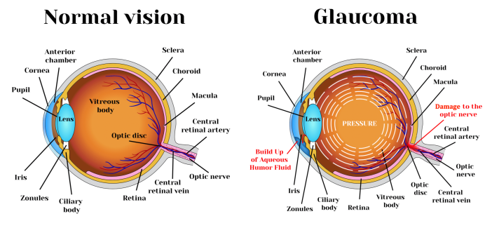
Glaucoma is a buildup of pressure within the eye that causes damage to the optic nerve. Glaucoma can occur at any age in a person with Aniridia, but most commonly begins in childhood or early adulthood. About 50 percent of people with Aniridia develop glaucoma by age 8. The risk for glaucoma increases with age.
Normally, the eye is filled with a fluid called the aqueous humor. This fluid gives the eye its round shape and is constantly being produced and drained, keeping pressure inside the eye in balance.
Glaucoma occurs when the fluid does not flow out as easily as it flows in. When too much fluid builds up inside the eye, it causes intraocular pressure, or IOP, to go up. When the IOP is too high for too long, it puts pressure on the inner walls of the eye, eventually causing damage to the optic nerve. The optic nerve is responsible for transmitting visual signals from the retina to the brain. Damage to this nerve can lead to vision loss and irreversible blindness.
People with Aniridia are at increased risk for development of glaucoma because of abnormalities in the eye that can disrupt drainage of the aqueous humor.
Treatment of glaucoma in patients with Aniridia can be complex and challenging but most patients can be successfully treated. Referral to a knowledgeable and experienced specialist is important.
Early detection and appropriate treatment of glaucoma can help to preserve vision in patients with Aniridia. Recent developments in medical and surgical treatment are promising.
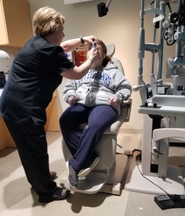
Jenna undergoing eye exam
Assessment and diagnosis of glaucoma may involve some or all of the following tests:
Tonometry measures the eye's intraocular pressure or IOP. The doctor uses anesthetic eyedrops to numb the surface of the eye, then places a special device directly on the cornea. "Applanation tonometry," the "Tonopen" and the "Rebound Tonometer" are common devices for measuring intraocular pressure. These devices provide a more accurate measurement than the “puff of air” -type machines, which are typically used for routine screening. Applanation tonometry is the most accurate method, but the Tonopen and Rebound Tonometer may also be used, especially for children.
NOTE: Normal IOP ranges from 12-22 mm Hg. However, common features of aniridic eyes such as nystagmus and increased corneal thickness can make accurate measurement of IOP challenging. Multiple readings taken over a period of time may be more helpful than a single measurement.
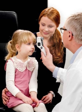
Rebound Tonometer
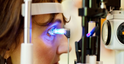
Applanation tonometry
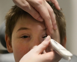
Tonopen
Gonioscopy is an eye exam using a special microscope called a slit lamp and a prism called a goniolens to look at the anterior chamber angle at the front of the eye. The "angle" is where the cornea and the iris meet, and where the aqueous humor drains. In people with Aniridia, the angle may be abnormal or may not function properly.
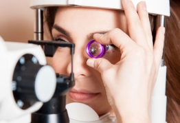
Gonioscopy
Ophthalmoscopy is used to examine the inside of the eye, especially the optic nerve and optic discs. In a darkened room, the doctor will look at the eye with an ophthalmoscope (an instrument with magnifying lenses and a small light). This instrument allows the doctor see the size, shape and color of the optic nerve, as well as other structures at the back of the eye.

Ophthalmoscopy
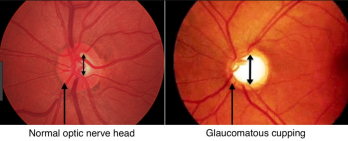
What the doctor sees through the ophthalmoscope
Glaucoma in patients with Aniridia is generally treated with medication or surgery or both. Eye drop medications work by decreasing the amount of fluid produced by the eye or by helping fluid to drain more effectively. Surgery may also improve drainage of fluid from the eye.
Eyedrops for treatment of glaucoma come in many different types, strengths, and combinations.
The physician will start by prescribing the smallest amount of medication that offers the best chance of controlling glaucoma, with the fewest side effects. Glaucoma medications must be taken daily, on a regular basis to control pressure in the eye.
Most glaucoma medications have some side effects, but these side effects may lessen after a few weeks. If a drug/dosage does not seem to be controlling glaucoma or when there are unacceptable side effects, a different drug or combination of drugs may be tried.
More information and a comprehensive list of medications used to treat glaucoma, along with mechanisms of action and side effects, is available.
Surgery
When medication does not control glaucoma or becomes ineffective, surgery may be necessary. Types of glaucoma surgery include the following:
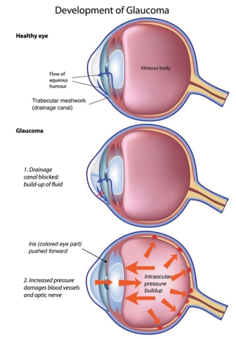
Trabecular meshwork
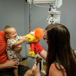
Infant Eye Exam
Measuring eye pressure in young children is often difficult because they cannot cooperate with the examination. If a child is crying when his eye pressure is measured, the reading may be artificially increased and will not be an accurate assessment.
Some physicians suggest using a mild sedative such as chloral hydrate, which may allow the child to be examined in the physician’s office. However, sedatives in children can have unpredictable effects and may not provide enough sedation at the right time to enable an examination.
Another option for obtaining accurate pressure readings in children is an Exam Under Anesthesia or EUA. An EUA is an examination conducted while the patient is under general anesthesia. Unlike a surgical procedure, an EUA requires a very small amount of anesthesia, for a short period of time. An important benefit of examining a child’s eyes under anesthesia is that it allows the physician to conduct a more thorough examination of the eye.
"Glaucoma in Aniridia - Part 1"
"Glaucoma in Aniridia - Part 2"
Sign up for News & Events
COPYRIGHT© 2025 IWSA / International WAGR Syndrome Association
